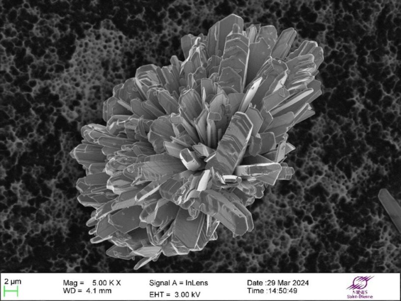
The Jacquet Prize recognizes the most beautiful images of materials (micro or macro-graphs) obtained using microscopic techniques. This year, 45 participants competed, and Maelig Ollivier, assistant lecturer at the SPIN center, won first prize.
The image is impressive; Maelig named it Tenorite Chrysanthemum because its shape reminded him of a flower. It shows the morphology of a copper monoxide powder grain observed under a scanning electron microscope. We can see that, far from being smooth and spherical, this powder grain has a complex morphology.
To better understand, we asked the laureate to describe the method used to capture this image. Here is his response:
“I used one of the school’s scanning electron microscopes. Like all microscopes, it allows us to visualize small objects that are not easily seen with the naked eye. The big difference from an optical microscope (magnifying glass, binocular, those we used in middle school) is the resolution: since we don’t use light but electrons for observation, we can obtain images where the smallest distinguishable details are a few tens of nanometers, 100 million times smaller than a meter. However, we inevitably get a grayscale image, regardless of what we observe.”
What is also noticeable about this Tenorite “flower” is the large surface area it has in contact with its environment. This has consequences for its reactivity, and it is this reactivity that Maelig Ollivier studies daily at the center for Industrial and Natural Process Science.